PHILADELPHIA – Last year, Thomas Jefferson University researchers built the first 3D map of the heart’s “brain” in rats. They identified the precise locations where the neurons are located in the heart and what their molecular properties are. Now, building on the same techniques, the group has demonstrated major differences between the heart’s little brain in male and female animals, with potential implications for human disease.
“We know that the symptoms of a heart attack are different in men vs. women – our hearts appear to respond differently to stress,” says co-senior author Rajanikanth Vadigepalli, PhD, professor of Pathology, Cell Biology and Anatomy Thomas Jefferson University. “Our studies provide new data towards figuring out true explanations for that observation. The little brains in the male and female hearts are localized and wired differently. The 3D map of the heart’s neurons in rats demonstrates some of those differences.”
The results were published in the Cell Press journal iScience on July 19th.
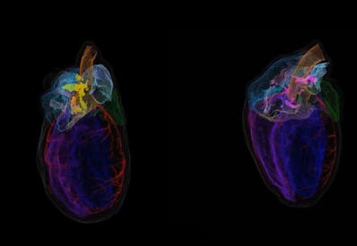
As part of a national NIH collaboration called SPARC that aims to understand how the nerves interact with each of the major organs in the body and influence their activity, the Jefferson team collaborated with Auckland Bioengineering Institute researchers to build a 3D digital scaffold of the rodent heart. That scaffold allowed the group to overlay data in a precise yet reproducible way across subjects as they collected thousands of images using high-throughput automated methods of tissues slices and imaging and then painstakingly marking the locations of neurons. The Jefferson group also collaborated with Dr. Zixi (Jack) Cheng’s group at the University of Central Florida and Strateos and MBF Bioscience to develop and annotate the cardiac feature a large collection of images at very high resolution of entire rat hearts. With that data, they built a 3D representation and image. The collaborative group compared the results from four male and three female rat hearts.
“This approach gave us a way to look at each individual heart on the single-cell level, and then compare across male and female rats hearts in a standardized way using unified 3D coordinates,” says co-senior author James S Schwaber, PhD.
The research team observed a good deal of variation between each individual animal irrespective of sex, even as the overall organization of the heart’s little brain showed conserved features across individual animals and sexes. “But the differences between male and female rats was profound and surprising,” says Ms. Shaina Robinson, a research assistant on the project, who poured over the images one by one to validate the annotation and developed visualizations that allowed systematic comparisons.
In general, female rats had far fewer neurons dappled across the heart (see video). “Although we could see that all of the essential functions were there, the hearts of female rats were able to carry out essential functions of the heart’s little brain with fewer numbers of nerve cells,” says Dr. Schwaber.
“Ultimately, we’re looking for a better understanding of heart disease and heart failure, which tend to be related to the nerve cells that are distributed in many places on the heart and control how the heart pumps at various speeds or strengths,” says Dr. Vadigepalli. “This sets the stage for answering new questions about human disease, in particular on sex differences in cardiovascular health, that we really haven’t had the comprehensive anatomical and molecular tools to answer before.”
For example, in a separate study, also publishing in iScience, the team explored the molecular properties of these neurons as they control heart function in pigs. They found that unlike neurons of the brain, which are often defined by the neurotransmitter they release – ones that activate or depress activity etc. – neurons of the heart had much more multifunctional capacity for neurotransmitters and modulators. In the heart, the neurons are engaged in a complex network of these modulators and their receptors that can extensively fine tune how the little brain at the heart processes the signals from the brain and controls the heart functions accordingly.
“This work demonstrates that the local control of the heart is much more nuanced than we’ve appreciated to date,” says Dr. Vadigepalli. “It suggests, for example, that a single drug that turns heartbeats up or down, may not be as effective long-term as one that hits the right notes, or combination of targets within this complex network. There is much more to explore, discover and translate for developing new therapies for heart disease.”
The work was driven by three research trainees: Shaina Robbins, a research assistant, and Alison Moss, a graduate student from Jefferson, and Clara Leung, a graduate student from the lab of the another co-senior author, Zixi (Jack) Cheng at the University of Central Florida.
Article references:
Alison Moss, Shaina Robbins, Sirisha Achanta, Lakshmi Kuttippurathu, Scott Turick, Sean Nieves, Peter Hanna, Elizabeth H. Smith, Donald B. Hoover, Jin Chen, Zixi (Jack) Cheng, Jeffrey L. Ardell, Kalyanam Shivkumar, James S. Schwaber, Rajanikanth Vadigepalli, “A single cell transcriptomics map of paracrine networks in the intrinsic cardiac nervous System,” iScience, DOI: 10.1016/j.isci.2021.102713, 2021.
Clara Leung, Shaina Robbins, Alison Moss, Maci Heal, Mahyar Osanlouy, Richard Christie, Navid Farahani, Corey Monteith, Jin Chen, Peter Hunter, Susan Tappan, Rajanikanth Vadigepalli, Zixi (Jack) Cheng, James S. Schwaber, “3D single cell scale anatomical map of sex-dependent variability of the rat intrinsic cardiac nervous system,” iScience, DOI: 10.1016/j.isci.2021.102795, 2021.
Media Contact: Edyta Zielinska, 267-234-3553, edyta.zielinska@jefferson.edu.