Human cells contain two distinct genomes, both of which provide key sets of instructions. A great deal is known about how our three billion base pair nuclear genome is transcribed into RNA, and how many of these RNAs are translated into proteins. In contrast, far less is understood about how the relatively minuscule sixteen thousand base pair genome in human mitochondria is transcribed. Understanding the molecular details of this mitochondrial process has important implications for a variety of human diseases, including developmental disorders, neurodegeneration, and cancer.
In a recent study published in Molecular Cell, Herbine et al. present high-resolution cryo-EM structures that capture key intermediates of transcription initiation in human mitochondria. These structures reveal how mitochondrial RNA polymerase (mtRNAP) assembles with transcription factors TFAM and TFB2M on the light-strand promoter (LSP), offering the first detailed view of promoter recognition, transcription bubble formation, and the roles of TFAM and TFB2M in positioning and activating the polymerase. The study reports eight novel conformations of transcription complexes, spanning the earliest steps of initiation through the transition to elongation, providing an unprecedented structural timeline of mitochondrial transcription.
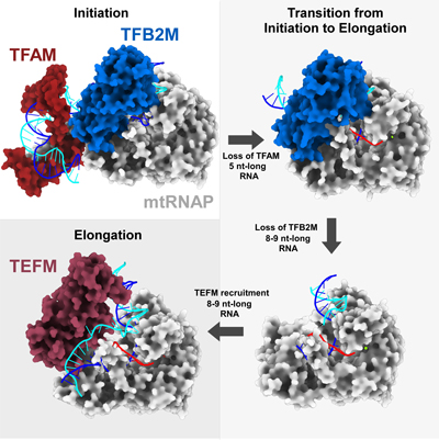
Using state-of-the-art single particle analysis, the authors determined — for the first time — the structures of initiation and elongation complexes of mitochondrial RNA polymerase (POLRMT) originating from a natural, fully double-stranded promoter. This breakthrough opens a new direction in the mitochondrial transcription field, allowing researchers to study transcription intermediates in their native environment. The work further uncovers a stepwise mechanism by which TFB2M disengages from mtRNAP and DNA, enabling the transition from initiation to elongation. Structural rearrangements in the DNA template and RNA product disrupt interactions with TFAM and TFB2M, allowing processive RNA synthesis to begin. These findings significantly advance our understanding of mitochondrial gene expression and lay the groundwork for future studies on transcription regulation and mitochondrial disease mechanisms.