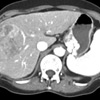
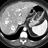
CT demonstrates a large hepatocellular carcinoma in a 60 year old male with a history of hepatitis C. His alpha fetoprotein was 12,000 IU/Liter

Interventional Radiology


Chemoembolization
Chemoembolization is a technique that has been very successful at treating primary liver tumors (hepatocellular carcinoma) or biologically active tumors that have spread to the liver (carcinoid or neuroendocrine tumors). This procedure involves delivering high doses of chemotherapy to the tumors while cutting off their blood supply. This limits the effect on the surrounding healthy parts of the liver. Patients also avoid the adverse effects of chemotherapy, such as hair loss and diarrhea. A catheter is placed into the artery in the groin and is passed into the artery to the liver. Chemotherapeutic drugs and particles to block flow are infused into the tumor bed. Patients usually stay one night in the hospital following this procedure. The Interventional Radiology physicians at Thomas Jefferson perform over 500 chemoembolization procedures/year and have published extensively on the topic.
Case Study Chemoembolization of Hepatocellular Carcinoma:
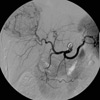
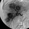
Arteriogram outlines the supply to the tumor (B)
as well as the hypervascular blush (C)
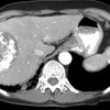
One month following chemoembolization,
no residual enhancement is identified.
The alpha fetoprotein is 70 IU/Liter.
Case Study Chemoembolization of Neuroendocrine/Carcinoid Tumor:
This 53 year old male had several episodes of watery diarrhea daily which was refractory to somatostatin administration. Angiography demonstrates multiple hypervascular metastases in the liver.
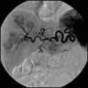
The right hepatic artery is selected.
Treatment was performed from this location.
Within two days, the patient’s symptoms had completely stopped. CT scan performed a month after chemoembolization demonstrates dense uptake of the chemoembolic material within the tumors.