Interventional Radiology

Vascular Malformations

Vascular malformations are areas of abnormal vascular tissue that can be focal or diffuse and can arise anywhere in the body. They are caused by an error in the development of blood or lymphatic vessels. They are present at birth but may not become apparent until later in childhood, adolescence or adulthood. Vascular Malformations generally grow in proportional to overall growth although they may expand rapidly in response to adolescence, pregnancy or other hormonal changes. They never involute or regress spontaneously. There are several different types of vascular malformation. Most occur sporadically but some forms can be inherited. They can be made up of arteries, veins, capillaries, or lymphatic vessels and they are named according to which type of vessel is predominantly affected.

Treatment for vascular malformations depends upon the type. Each type of malformation is treated differently. AVMs are usually treated by radiologically guided injection of an embolic agent or sclerosant into the abnormal vessels within and supplying the malformation. VMs are usually treated by radiologically guided direct injection of sclerosant which obliterates the abnormal venous channels. LMs are also treated by direct injection of different types of sclerosants. Often, a combination approach is needed for effective treatment of the malformation.

Venous Malformation

VMs are the most common type of symptomatic vascular malformation. They can be single or multiple. They most commonly involve the skin but can involve any tissue or organ in the body. Rarely, they can occur as part of more generalized syndromes such as Blue Rubber Bleb Nevus Syndrome or Maffuci’s Syndrome. VM’s are composed of abnormally formed, dilated veins with very thin walls due to a relative lack of smooth muscle cells. VM’s are often mistakenly called hemangiomas. While present at birth, VM’s may not be obvious and may not ‘appear’ until later in childhood or adulthood. When they involve skin they are usually soft, compressible dark blue bulges. If they become hard and painful, this usually indicates blood clot formation within the VM and expert consultation should be sought. VMs can enlarge in response to injury, puberty, pregnancy or the oral contraceptive pill (OCP). Large VMs can bleed spontaneously or can be associated with bleeding and clotting abnormalities due to consumption of platelets and clotting factors.

VMs are treated with sclerotherapy or surgical debulking. Surgery is more invasive than sclerotherapy and is rarely curative. Sclerotherapy involves injecting an irritant chemical into the abnormal venous channels, usually under image guidance, in order to promote obliteration of the venous channels and shrinkage of the VM.

Arteriovenous Malformation

AVMs are composed of congenital abnormal communications between arteries and veins. They most commonly involve the brain but can also involve the head & neck, the limbs, trunk and internal organs. The microscopic capillary bed which normally intervenes between arteries and veins is absent resulting in higher than normal blood flow. AVMs can be sporadic or can occur in conditions such as Hereditary Hemorrhagic Telangiectasia (HHT or Osler-Weber-Rendu Syndrome), Parkes-Weber Syndrome, Cobb Syndrome or in association with PTEN genetic mutations.

AVMs involving the skin and superficial tissues present with gradual enlargement and color changes. Local warmth associated with an enlarging pulsatile mass is a common presentation. Progression with pain, ulceration or bleeding is not uncommon. With time, blood flow through an AVM can become so fast that the heart can become overworked.

Whenever an AVM is suspected, expert consultation should be sought. AVMs can be treated by angiographically guided endovascular and percutaneous embolization and sclerotherapy. This involves delivering the embolic/sclerosant material to the AVM either through a catheter inserted through an artery in the groin or through a needle placed directly through the skin. Surgical resection can sometimes be ‘curative’ for suitable small focal AVMs.

Lymphatic Malformation (LM)

LMs are sponge-like collections of lymphatic channels and cystic spaces that contain clear lymphatic fluid. LMs can be macrocystic or microcystic depending on whether they contain large or microscopic fluid spaces. They most commonly occur in the head & neck and upper chest but can occur in any location throughout the body. They usually present as localized swellings which can worsen with a generalized viral or bacterial illness. Infection and bleeding within an LM can cause periodic worsening of symptoms. Depending on the location of the LM, leakage of lymphatic fluid may cause excess fluid in the abdomen or chest (chylous ascites or chylothorax) and generalized protein loss. Treatment can either be surgical excision for small focal lesions or sclerotherapy which involves directly injecting the LM with an irritant chemical that results in sclerosis and regression over a short period of time.

Capillary Malformation (LM)

CMs generally present after birth. CMs are flat, sharply demarcated, red-pink staining of the skin, sometimes referred to as a "port-wine stain". CM’s most commonly involving the head and neck. Most are harmless birthmarks but they can signal an underlying abnormality if they involve the upper eyelid, forehead or overly the spinal column. They are often present as part of combined vascular malformations associated with limb overgrowth. CMs are treated with pulsed-dye laser and are generally not treated by Interventional Radiology techniques.

Combined Vascular Malformations

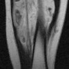
Combined malformations affect the extremities and trunk and are associated with limb overgrowth in both girth and in length. Combined malformations involve more than one tissue/channel type. They can be accompanied by overgrowth of soft tissue and bone in the affected region/extremity. They sometimes affect more than one extremity and often involve the perineum and trunk. The most common forms are Klippel-Trenaunay Syndrome (KTS) and Parkes-Weber Syndrome (PWS).

KTS is a combined Capillary-Lymphaticovenous Malformation (CLVM), characterized by capillary staining, venous anomalies of the deep and superficial systems, lymphatic malformations and limb overgrowth involving soft tissue and bone. KTS usually involves the lower extremity and trunk in combination but can also involve an upper extremity. The lymphatic component of the syndrome is prone to infection and internal bleeding while the venous component can be complicated by blood clot formation which can migrate to the lungs.

PWS is a combined Capillary-Arteriovenous Malformation (CAVM), characterized by capillary staining, multiple AVMs and overgrowth of the affected extremity. The AVM component of this syndrome is most troublesome with pain, skin breakdown, bleeding and often high output cardiac failure due to the number and size of the arteriovenous shunts.

Treatment of combined malformations includes interventional radiological techniques discussed previously. A multi-disciplinary approach with input form Surgeons, Dermatologists, Hematologists, Radiologists and other specialties, is vital in managing patients with combined vascular malformations.

Sclerotherapy/Embolization Procedures

Treatment is preceded by an outpatient consultation at which time the patient is fully evaluated, the procedure is explained in full and any additional blood tests or radiological examinations are requested.

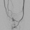
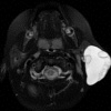
Radiologically guided treatment for all types of vascular malformation involves percutaneous (through the skin) and endovascular (through blood vessels) placement of needles and catheters to deliver the sclerosant or embolic material to the affected lymphatic or vascular channel. Chemical agents used include Sodium Tetradecyl Sulfate (STS), Alcohol, n-Butyl Cyanoacrylate (n-BCA), Onyx ® liquid embolic agent, Doxycycline and Bleomycin. Other agents used include endovascular coils and endovenous laser.

Delivery of sclerosant can be painful and procedures can be lengthy. For both these reasons, it is preferable to perform sclerotherapy under general anesthesia. Treatment is usually followed by admission for observation with patients normally discharged the day following the procedure.

To arrange a consultation or if further information is required, please contact CVIR (Cardiovascular Interventional Radiology) at: 215-955-6440