Interventional Radiology

Uterine Fibroids & Uterine Artery Embolization

What are uterine fibroids?

Uterine fibroids (leiomyomas or myomas) are the most common benign (non-cancerous) tumor of the female reproductive tract.  Up to 40% of women age 35 and older and up to 50% of African American women have uterine fibroids.

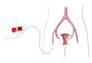
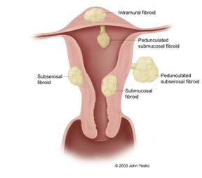
Fibroids may be located within the wall of the uterus (intramural), on the surface of the uterus (subserosal), or beneath the lining of the uterine cavity (submucosal) (Figure 1 to the right).  Some fibroids are located on a stalk and are referred to as pedunculated fibroids.

What are some of the symptoms associated with uterine fibroids?

Symptoms associated with uterine fibroids include prolonged, heavy menstrual cycles or menorrhagia, dysmenorrhagia (abnormal bleeding between menstrual cycles), fatigue from iron-deficiency anemia (low blood count), pelvic/back pain, constipation, obstipation (painful bowel movements), dyspareunia (painful sexual intercourse), bloating, and urinary frequency.

How are fibroids diagnosed?

Fibroids are diagnosed by physical exam and by imaging techniques such as ultrasonography (Figure 2, to the left) or magnetic resonance imaging (MRI) (Figure 3, on right).

What are the treatment options for uterine fibroids?

There are several treatment options for symptomatic uterine fibroids.  The most conservative treatment is medical management.   This includes the use of medications to control pain and cramping such as nonsteroidal anti-inflammatory drugs (i.e. Motrin).  Birth control pills and other hormonal therapy (i.e. Lupron) may also be used to control symptoms however; certain hormones produce side effects and can only be used on a temporary basis.

Hysterectomy (surgical removal of the uterus) and myomectomy (surgical removal of visible fibroids) are the most familiar treatment options for uterine fibroids. 

Hysterectomy is usually performed as an open surgical procedure.  The procedure requires general anesthesia and the recovery time is approximately 6 weeks.  However, some hysterectomies can be performed laparoscopically allowing for a shorter recovery time. 

There are three different types of myomectomy: hysteroscopic myomectomy, laparoscopic myomectomy and abdominal myomectomy.  All three require general anesthesia.  Hysteroscopic myomectomy requires the shortest recovery time but is reserved for fibroids located within the uterine cavity or just under the uterine lining.  While myomectomy is often successful in relieving symptoms related to fibroids, the more fibroids a woman has, the less successful the procedure.

What is fibroid or uterine artery embolization?

Uterine artery embolization (UAE) is a favorable treatment alternative for women with fibroids who want to preserve their uterus and avoid surgery.

UAE is a minimally invasive procedure performed by an interventional radiologist through a tiny nick in the skin while the patient is conscious but sedated.

The interventional radiologist inserts a catheter (hollow tube) into an artery located in the groin.  Under fluoroscopic (X-ray) guidance, the catheter is advanced into the vessels (uterine arteries) supplying blood flow to the uterine fibroids (Figure 4 to the right).

Once the catheter is positioned in the uterine arteries, small particles (Figure 5, to the left) are injected to block blood flow to the uterine fibroids.  

Following embolization of the uterine arteries, the catheter is removed and compression of the catheter insertion site is performed for approximately 15 minutes.  The patient is required to lie flat for 6 hours following the procedure.  A majority of patients are discharged the same day as the procedure or the following morning. 

What are the complications associated with uterine artery embolization? 

Although UAE is considered a very safe treatment, there are a few complications associated with the procedure.  Infection occurs in a small number of patients and is usually treated with a course of oral antibiotics.  Injury to the uterus potentially leading to hysterectomy is fortunately rare (<1% of patients).  Women following the procedure may develop fever, pain and nausea, which are managed with pain medications, anti-nausea medications and Tylenol.   Premature menopause is another potential procedural risk. However this is unusual (3%) in patient less than 45 years of age. 

Despite the complications mentioned above, complications associated with UAE are lower than those related to the surgical alternatives; myomectomy and hysterectomy. 

How successful is uterine artery embolization?

Fibroids typically decrease by 50-60% of their original size after UAE (Figure 6).  The uterus also decreases in size by approximately 50% of its original size (Figure 7).  As far as symptom relief, success rates are 90% or greater for heavy bleeding and 85% or greater for bulk-related symptoms (i.e. urinary frequency). 

Figure 6:  Pre-UAE (left) and three months post-UAE (right) pelvic MRI.   Large fibroid (black circle on right) is smaller in size and no longer has blood supply.

Figure 7:  Pre-UAE (left) and three months post-UAE (right) pelvic MRI.  Two fibroids (black circles, right) no longer have blood supply and the uterus is smaller. 

How do I determine if I am a candidate for UAE?

Call Interventional Radiology at 215-955-6440 to schedule a consultation.  Our interventional radiologists will help you determine if UAE is right for you.