Ultrasound

Services Offered

Diagnostic ultrasound imaging, or sonography, has progressed dramatically over the last decade. The diagnostic utility of the modality is higher than ever before and examinations can generally be performed more rapidly without sacrificing accuracy. In addition to the most common clinical applications, our staff has developed a wide range of new uses of ultrasound which has lead to a more diverse spectrum of diagnostic options. Jefferson’s Division of Diagnostic Ultrasound maintains nationally recognized accreditation in all aspects of ultrasound imaging, including general radiology, obstetrics and gynecology as well as non-invasive vascular imaging.

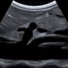
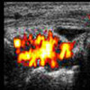
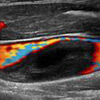
Three-dimensional Sonohysterography Demonstrates a Septate Uterus with Polyps

The use of ultrasound imaging in obstetrics and gynecology is well established and our center continues to stay at the leading edge in these areas. In the recent past we have developed and utilized sophisticated techniques such as three-dimensional sonohysterography to provide our patients with the highest level of services.

Ultrasound Imaging of Normal Aorta and its Branches

Abdominal applications of ultrasound imaging continue to provide valuable contributions to medical diagnostics. The use of sonography to evaluate such structures as the liver, gallbladder, kidneys and pancreas has proven invaluable for patient management and the modality is recognized as an important screening tool for aortic aneurysms. Utilizing the latest technological advances in instrumentation allows the detection of very subtle organ abnormalities and the highest degree of diagnostic confidence.

Musculoskeletal Imaging

One of the fastest growing areas of ultrasound is Musculoskeletal Imaging. A wide-range of sports, ergonomic, or other injuries can be detected with the use of high frequency imaging. Our staff is pioneering new minimally-invasive ultrasound-guided therapies which are rapidly changing how patients are treated and speeding their recovery process.

Color Doppler Imaging Demonstrates a Deep-venous Thrombosis

Our vascular laboratory is internationally-recognized in this diagnostic specialty area. Non-invasive vascular studies of all types are performed by certified vascular technologists with interpretations provided by highly trained radiologists. Our imaging professionals work closely with vascular surgeons and members of other medical disciplines to enhance patient care. The most common applications of vascular ultrasound include evaluation of the cerebrovascular system for stenoses and peripheral veins for the presence of thromoses (blood clots).

To schedule an examination call 1 800-JEFF NOW