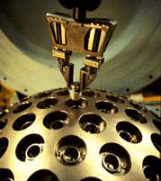
Our unit was installed and became operational in June of 1996, and we have treated over 550 patients with intracranial lesions or functional disorders on this unit. Historically, the Gamma Knife represents the first radiosurgery instrument developed originally in Sweden by Dr. Lars Leksell in 1950. Based on the principle of precise intracranial localization utilizing a cartesian coordinate system, Dr. Leksell designed a fixed hemisperical array of Cobalt sources which emmited gamma rays to one precise point in space.

Jefferson Brain Tumor Center
Contact Us
Name:
Jefferson Brain Tumor Center
Academic Offices
901 Walnut Street, 3rd Floor
Philadelphia, PA 19107
Contact Number(s):
Make an Appointment
New Patients - 215-503-6987
All Other Patients - 215-955-7000
The Radiosurgery Units operate in the Jefferson Hospital for Neuroscience, and both Units are co-located with an MRI scanner, a CAT scanner, a neuroangiography suite, and a neuroradiology reading room, providing a complete integration of all the technology and staff support necessary for the practice of radiosurgery. The Radiosurgery Units are comprised of a U-model Leksell Gamma Knife and a Varian 600SR LINAC designed for and dedicated to radiosurgery. Each of these units are decribed in detail below.
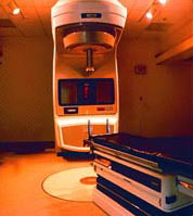
Our unit was installed and became operational in October of 1994, and we have treated over 600 patients with intracranial lesions on this unit. This unit represents its first world commercial installation based on a prototype developed by the Varian Corporation in collaboration with the Joint Center for Radiation Therapy at Harvard University and the Radionics Corporation.
In the case of radiosurgery utilizing X-Knife, mastery of radiobiological principles must precede the practice of radiosurgery since any one of an array of techniques ranging from single fraction SRS to conventional fraction SRT might be best suited for an intracranial lesion. Since radiobiological models, for example, ascribe a direct relationship between late normal tissue damage and dose per treatment delivered to these tissues 10, 11, recently published series from a growing number of institutions, including our own, have explored the use of LINAC-based SRT for the treatment of benign tumors such as acoustic tumors. Although this experience is smaller and more recent, SRT data reflect comparable tumor control rates and higher rates of cranial nerve preservation, most notably hearing preservation 12-14.
At our institution, we have established a stereotactic radiosurgery program that includes the use of a LINAC designed for and dedicated to SRS and SRT, the Varian 600SR15. Recognizing that most institutions have general-purpose LINACS, we will describe our experience utilizing both techniques, which should be generally applicable to the typical practice of SRS and SRT, and include case treatments as examples of each.
The Jefferson Neurosurgical OR suites are equipped with the latest technology and dedicated Neurosurgery OR personnel to enhance the effectiveness and safety of neurosurgical procedures.
Neurosurgical equipment and personnel for brain tumors
- Zeiss Varioscope Operating Microscope
- Codman Rigid Neuroendoscopic (Zamarano neuroendoscope)
- Radionics OTS image guidance system
- Grass stimulator/ Nicolet Neurophysiology workstation
- Midas Rex Dissection System
- Dr. Paul Adou, dedicated Neuroanesthesiologist
- Dr. Daniel Shwartz and Surgical Associates (intraoperative neurophysiologic monitoring of brain stem and cranial nerve function)
- Dr. Michael Sperling and Associates (intraoperative corticography)
- Tina Nimmons, RN & Darryl Worrel, dedicated Neurosurgical OR staff