Breast Imaging

Services Offered

Screening breast imaging for early cancer detection.

Screening Mammography is the Cornerstone of Breast Cancer Screening

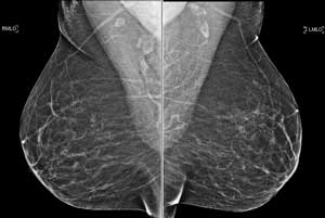
A screening mammogram is an X-ray of the breast used to detect breast changes in women who have no signs or symptoms of a breast abnormality or problem. A regular schedule of screening mammograms offers women the best way of detecting cancerous and pre-cancerous conditions early, when treatment can be most effective. At Jefferson, all mammograms are performed using digital breast tomosynthesis, which allows for more detailed evaluation of breast tissue and results in improved cancer detection while reducing unnecessary (non-cancer) imaging and biopsies.

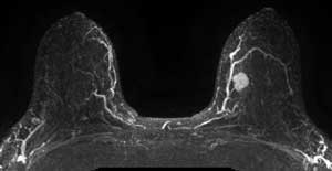
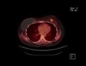
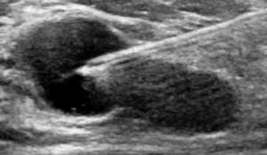
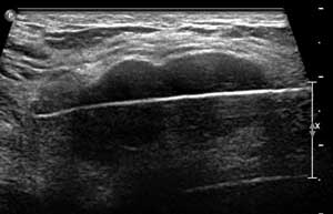
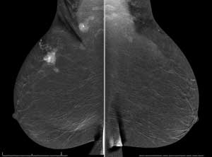
Supplemental screening technologies may benefit certain women in whom standard mammography may not be sufficient for cancer screening. These include women with dense breast tissue on mammogram, which may obscure small cancers, and women at a higher-than-average risk for breast cancer due to strong family history of breast cancer or genetic predisposition to breast cancer. These supplemental screening technologies include whole-breast (complete) screening ultrasound (hand-held or automated), MRI, and contrast-enhanced mammography.


Diagnostic breast imaging for problem solving

Diagnostic Mammography

A diagnostic mammogram is an X-ray of the breast that is performed to evaluate a specific problem or symptom in the breast. Diagnostic mammograms may be used to evaluate abnormal findings on a screening mammogram, or for women who feel a lump or other breast abnormality. 

Contrast-enhanced Mammography 

Targeted breast ultrasound

Contrast-enhanced mammography (or CEM) is a diagnostic mammogram where breast images are taken after administration of iodinated contrast dye through the vein. The addition of contrast helps find cancer because it highlights tumorous vessels and “contrasts” tumors against the background of normal breast tissue.

Breast ultrasound is a safe, noninvasive test that uses sound waves to image the inside of the breast. Targeted (or limited) ultrasound is used to help evaluate a specific physical breast problem such as a lump or a specific problem finding on another imaging test such as mammogram or breast MRI.

Breast MRI

Breast MRI (Magnetic resonance imaging) uses a powerful magnetic field, radio waves and a computer to produce images of the breast. MRI of the breast is not a replacement for mammography or ultrasound but rather a supplemental tool for detecting and staging breast cancer and other breast abnormalities. Breast MRIs may be helpful in certain women who are at high risk for developing breast cancer in their lifetime and may have value for women with breast cancer.

Whole-body PET/CT

Advanced breast cancer is a systemic disease and therefore we utilize PET/CT to provide fused whole body metabolic and cross-sectional imaging to help stage the disease and follow response to chemotherapy.


Imaging-guided breast intervention

Imaging-guided Breast Core Needle Biopsy & Cyst Aspiration

Breast core needle biopsy is the standard of care for minimally invasive tissue diagnosis and an alternative to more invasive surgical biopsy. Small samples of tissue are removed from the breast using a hollow core needle with or without a vacuum-assisted device that is precisely directed to the correct location using guidance of various imaging modalities: ultrasound, digital breast tomosynthesis (stereotactic), contrast-enhanced mammography, and MRI.

Ultrasound-guided Breast Cryoablation for Fibroadenoma

Fibroadenoma is the most common non-cancerous tumor of the breast, sometimes causing a lump and pain. Cryoablation is an FDA-approved treatment for fibroadenomas and is an alternative to open surgery. It uses extreme cold to eliminate tumorous tissue. Women with one or more fibroadenoma tumors measuring at or under 4 cm may benefit from the procedure.

Cryoablation is an outpatient procedure performed in office by a breast imaging radiologist. The procedure takes less than 30 minutes, with local anesthetic. A probe is inserted into the breast, under ultrasound guidance, through a tiny incision. Liquid nitrogen circulates through the probe, causing a rapid temperature drop that freezes and destroys the breast tumor from the inside out.

Breast cryoablation for fibroadenoma is considered a comfortable procedure, with cold acting as a natural pain reliever. No general anesthesia or post-procedure prescription pain medicine is needed.

Because there is no tissue removal, patients should expect a speedy recovery. The procedure preserves breast volume and minimizes risk of bleeding or infection. Cryoablation can be safely repeated for multiple tumors with no change in breast appearance.

This treatment has been successfully and safely used for treatment of non-cancerous breast disease since 2001. The device used at Jefferson Health was approved by the FDA in 2010. Ninety percent of patients report good or excellent cosmetic results one year after treatment.

To schedule an examination call 1-800-JEFF NOW