Musculoskeletal Radiology Fellowship

Leadership

Name: Jeffrey Belair, MD
Position: Fellowship Director

Leadership

Name: Vishal Desai, MD
Position: Assistant Fellowship Director

For Program Information

Name: Isabella Corcoran
Position: Program Coordinator
Contact Number(s):

Visiting Fellowships - Ultrasound

Director: Levon N. Nazarian, MD

Tuition: $1,000/day, $4,800 per week, $3,600 for each additional week up to 1 month, $2,000 per week thereafter

Approximate number of cases per day: 15-20

The primary focus of this visiting fellowship is clinical diagnostic applications including the major joints, ligaments, tendons, muscles and nerves. A limited number of ultrasound-guided injections, dry needling and other interventional procedures are performed. Visiting fellows have the opportunity to participate in the daily functioning of the facility by attending reading sessions and observing procedures.  The visiting fellow also has access to our academic resources including a library of recorded classroom courses covering most aspects of diagnostic ultrasound.

This fellowship is designed to meet the following practice gap: there are very few centers in the country that perform a high volume of MSK Ultrasound, so there is a great need to educate clinicians as to the proper methods and interpretation of these studies. There are clinicians who lack exposure to ultrasound guided interventional techniques.

Learning Objectives

  • Describe  the indications and techniques for MSK US imaging.
  • Identify pathologic conditions seen on MSK US.
  • Discuss the MSK procedures that can be performed with US guidance.


Director: Praveen Mehotra, MD

Additional Faculty:  Norman Alexander, AAS, RDCS

Tuition: $1,000/day, $4,800 for the 1st week, $3,600 for each additional week up to one month, $2,000 per week thereafter. 

Approximate number of cases per day
: 30

Visiting fellows will participate in a comprehensive overview of clinical echocardiography as experienced during a typical day in our laboratory. Routine 2D/Doppler, stress (exercise and pharmacologic), and transesophageal echocardiograms are performed daily in our laboratory and will permit the program to be tailored to an individual’s needs. Fellows will observe studies being performed by sonographers and physicians. Daily interpretative reading sessions will allow the fellow one-on-one interaction with our staff echocardiographers. Advanced techniques such as 3D, contrast, and strain echocardiography are also performed regularly and exposure to these advanced analysis techniques can be arranged for advanced fellows.

This fellowship is designed to meet the following practice gap: the average practitioner performing ultrasound is not trained in echocardiography. There are practitioners who do not have exposure to 3D, contrast and strain echocardiography

Learning Objectives

  • Recognize the views and components of a standard 2D and Doppler echocardiogram.
  • Identify how to interpret a standard 2D and Doppler echocardiogram.
  • Practice the interpretation of additional echo modalities such as stress and transesophageal echocardiography.
  • Identify the indications and benefits of advanced echo techniques such as 3D, contrast, and strain echocardiography.


Director: Sharon Segal, DO

Tuition: $1,000 for 1 day, $500 for second and third day.

The Thyroid Biopsy tutorial is currently offered only on Wednesday each week.  This visiting fellowship provides observation of ultrasound scanning and ultrasound-guided biopsy in 6-10 cases per day.  The visiting fellow has extensive one-to-one interaction with Dr. Segal throughout the day. The fellowship includes a one-to-one didactic session (lecture) covering thyroid disease and the role of ultrasound imaging and biopsy in diagnosis and treatment. The visiting fellowship may be extended to a second or third day focusing on techniques of ultrasound-guided procedures and set-up of a dedicated ultrasound interventional procedure area. The volume of cases seen here will enable physicians to learn how to detect nodules and to obtain material for cytologic evaluation.

This fellowship is designed to meet the following practice gap: physical examination is inaccurate for detecting and characterizing thyroid nodules. Further tests are needed for an accurate diagnosis.  There are not enough practitioners that can accurately detect and biopsy non palpable nodules.

Learning Objectives

  • Identify the role of ultrasound imaging in thyroid disease.
  • Calculate an algorithm for recommending thyroid/neck FNA based on imaging results.
  • Observe the technique of ultrasound-guided FNA of thyroid/neck masses.
  • Learning objectives for the interventional procedures extended second and third days:
  • Recognize the role of ultrasound-guided interventional procedures.
  • Explain the process of setting up a new dedicated interventional service.


Director: Baskaran Sundaram, MD

Tuition: $1,000/day, $4,800 for the 1st week, $3,600 for each additional week up to one month, $2,000 per week thereafter. 

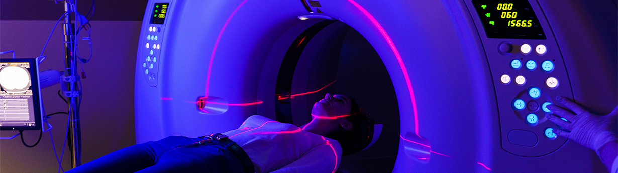
Every year, our department serves over half-a-million patients and enjoys high-level satisfaction grades from our patients and clinicians. Our department’s patient interactions include approximately 9000 non-invasive CT/MRI angiography exams and 1500 cardiac CT/MR exams,  performed for a range of clinical indications. Our scanning machines are regularly upgraded and maintained with tight quality control measures to ensure safety and quality. Our quality control includes radiation dose reduction and prevention for contrast-induced nephropathy, gadolinium-induced nephrogenic systemic fibrosis, and contrast reactions.

Our cardiothoracic radiologists are fellowship-trained and highly experienced faculty members. They provide comprehensive imaging interpretations with a strong passion for cardiothoracic imaging. Cumulatively, they have more than 50 years of cardiothoracic imaging experience that includes years of CT, MRI, nuclear medicine, echocardiography, and conventional angiography. They are nationally recognized experts with leadership roles in radiology societies. They have produced numerous cardiac and thoracic imaging-related scholarly products, including external presentations, book chapters, books, and peer-reviewed and non-peer-reviewed publications.

Our thoracic CT examinations indications include imaging for thoracic malignancies, indeterminate lung nodules, lung cancer screening, and interstitial lung disease. Our cardiovascular examinations include cardiac CT for coronary calcium scoring, coronary CT angiography, CTA for Transcatheter aortic valve replacement (TAVR) planning, CTA for robotic minimally invasive cardiac surgery, and pulmonary vein mapping. Our MRI examination indications are for the assessment for mediastinal masses, and cardiovascular MRI for ischemic and non-ischemic cardiomyopathy, valvular heart diseases, congenital heart diseases, and cardiac masses.

Our visiting fellows have the opportunity to review and update all aspects of cardiothoracic imaging, including the exam indications, scan parameters, optimizing scan techniques, 3D post-processing, and standardized reporting. We use high-speed CT scanners that have the ability for single heartbeat imaging, and 1.5 and 3 Tesla MRI machines. We also use modern imaging techniques for cardiothoracic imaging with digital radiography, dual-energy CT scanning, and 3T MRI. We continue to perform research and integrate recent advancements in cardiothoracic imaging, including the use of artificial/augmented intelligence.

We accommodate combining cardiothoracic visiting fellowship with other radiology specialties to suit the need of the visiting fellows. We look forward to having you as our visiting fellow in our department.

This fellowship is designed to meet the following practice gap: advanced cardiothoracic imaging techniques are not widely used in clinical practice.  Practitioners often do not know indicators and techniques for those studies.

Learning Objectives

  • Identify the updates in multimodality imaging, management guidelines and practice patterns in thoracic malignancies, indeterminate lung nodules, lung cancer screening, and interstitial lung disease.
  • Apply modern non-invasive cardiothoracic imaging with regards to the scan parameters, scan techniques, 3D post-processing, and standardized reporting and report templates of coronary calcium scoring, coronary CT angiography, CTA for Transcatheter aortic valve replacement (TAVR) planning, CTA for robotic minimally invasive cardiac surgery, dual-energy scanning and pulmonary vein mapping.
  • Apply cardiac MRI with regards to the scan parameters, scan techniques, 3D post-processing, and standardized reporting and report templates of Our MRI examination for the assessment for mediastinal masses, and cardiovascular MRI for ischemic and non-ischemic cardiomyopathy, valvular heart diseases, congenital heart diseases, and cardiac masses.